Anatomical Pathology
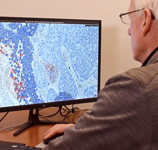
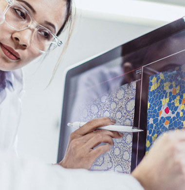
Flagship combines expert, board-certified pathologists with cutting-edge technology to get the most out of your clinical samples. To provide the actionable data drug developers seek, Flagship has developed one of the largest digital pathology and image analysis capabilities in the industry. Powered by our proprietary AI technology, this patented, cell-based tissue analysis technology can deliver high-complexity, data-rich tissue interpretations that remove the inherent variability of subjective manual scoring.
Digital Pathology Services
- Pathologist-directed image analysis
- Digital whole-slide brightfield and fluorescent scanning
- Multispectral imaging
- Dedicated team of imaging scientists, image analysts and application experts developing and implementing customized algorithms
- Flagship uses industry-standard scanning technology
-
- Chromogenic Brightfield: Leica Aperio AT Turbo
- Fluorescent: 3DHISTECH Pannoramic Scan II
- Multiplex Fluorescent: Akoya PhenoImager™ HT
-
Image Analysis
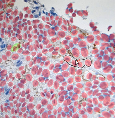
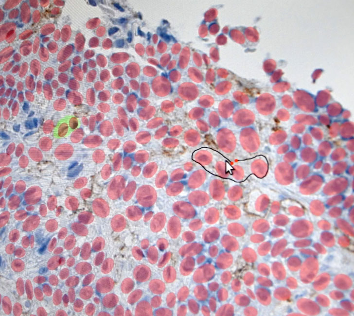
Flagship Biosciences has developed a proprietary, world-class image analysis platform called Flotilla. Flotilla is a flexible, multi-use image analysis platform optimized for use with clinical tissues. The program collects thousands of features per cell to create a cellular database for every sample. Machine learning is used to achieve tissue segmentation and cell identification.
IA Solutions
Flagship has established a library of validated algorithms to identify and quantify cells and biomarkers. These algorithms may be directly applied to your project, or customized as necessary.

